/ Dr. Silvia Maier
MRI – The technology
What is magnetic resonance imaging (MRI) and what happens during the investigation? Learn more about it from Dr. Silvia Maier (University of Zurich).
What is magnetic resonance imaging?
Magnetic resonance imaging (MRI) is an imaging procedure. It creates layer images of the inside of the body, which can be used to study the structure and function of organs and tissues.
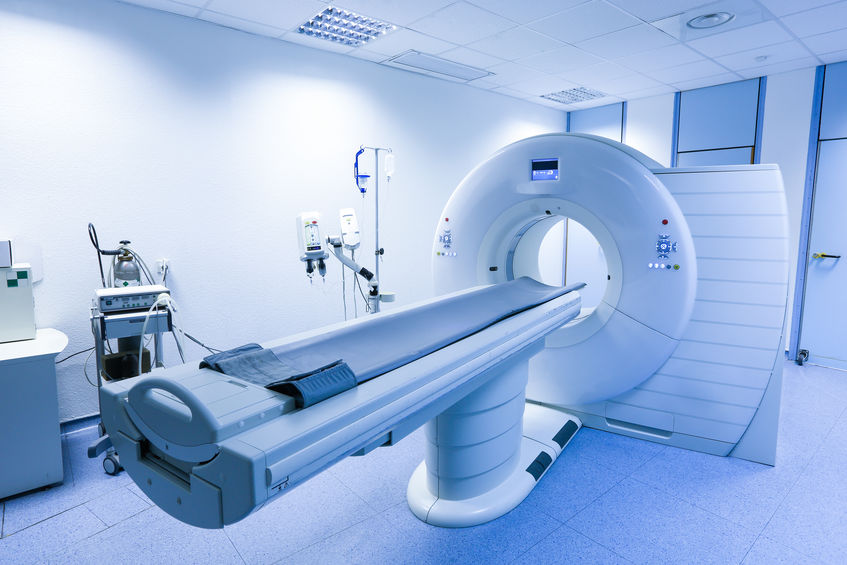
What happens during the investigation?
MRI works by means of a magnetic field in which the person to be examined is positioned. To collect data, the MRI scanner generates radio waves (similar to those used for radio and broadcasting) and additional magnetic fields that are much weaker than the basic field. The switching of these fields is characterized by loud knocking and humming noises. The hydrogen atoms in the body align themselves briefly with the additionally generated magnetic field before they align themselves again along the stronger basic field. The result is a signal that can be received by highly sensitive antennas and combined into an image by powerful computers.
Is something being administered to you?
No. In contrast to X-ray examinations or examinations with a computer tomograph (CT), MRI does not use X-rays. There is also no need for a contrast agent. The investigation is therefore harmless.