/ Nick Sidorenko, MSc
Shades of gray: what brain structure can tell about variability in risk attitude

In decision neuroscience, gray matter volume has been associated with risk preferences. Yet which brain structures are crucial for risk behavior and can we actually predict from just measuring a person’s brain how much risk they would take?
Gray matter matters
Taxi drivers have a lot of it in memory-related regions (1), professional musicians in the auditory cortex (2) and Einstein had 15% more of it in brain parts associated with mathematical thought (3). What, do you ask? Gray matter!
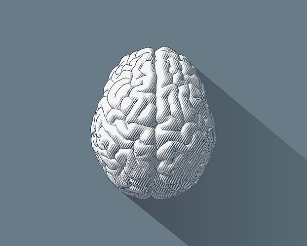
Gray matter represents about 40% of our brain and is composed of cell bodies of nerve cells (neurons). Their popular name little gray cells stems from the fact that they shimmer gray-ish when looked at with the bare eye. The more gray matter there is, the more neurons are available for processes as various as muscle control, sensory perception, memory, self-control, and last, but not least, decision-making.
Show me your brain and I can make a guess you how much you love risk
In decision neuroscience, gray matter volume has been associated with risk preferences. Yet which brain structures are crucial for risk behavior and whether we can actually predict from just measuring a person’s brain how much risk they would take is an open question. In one study, researchers from University College of London asked participants to choose between two options with different reward and risk levels (4). Then they scanned people’s brains using magnetic resonance imaging (MRI) and found that individuals with higher gray matter volume in the right posterior parietal cortex were more risk-tolerant while making their choices. The authors argued that this region, previously related to decision-making under uncertainty, might serve as a biomarker for financial risk attitude, if the results are replicated for a general population. Other scientific findings concerned a brain region called the insula. According to recent studies, the higher the financial risks taken during the task, the higher the gray matter volume in its anterior part (5), whereas the more loss averse humans are, the smaller the volume of its posterior part tends to be (6). Finally, in one of our recent posts, we talked about why older people may sometimes make poor financial decisions. Interestingly, economic irrationality in adults over 65 years old was also correlated with the gray matter volume, but this time in the ventrolateral prefrontal cortex (7).
But other life domains also provide risky choices. Differences in the gray matter volume of various brain regions have also been found to correlate with the risk of alcohol consumption (8, 9), cannabis use (10), and sensation-seeking behavior (11). In the digital world, internet gaming addiction (12) and even the number of friends on Facebook (13) were related to differences in volume of the brain regions.
Potential as a biomarker
Studies about gray matter provide promising results and open new horizons for better understanding of the neural basis of human risk behavior. But do they imply that we just need to find a way of boosting gray matter growth, for example, in our insula, to start investing in high-risk stocks? Actually… no. All these findings are of correlative nature and do not reveal what causes behavior we observe. To establish the causal relationship, decision neuroscientists use a range of other approaches to follow up on MRI findings with other tools of neuroscience.
What is interesting about gray matter is that its volume in different brain regions is relatively stable over long periods of time during adulthood. Thus, it holds potential as a biomarker, which could help scientists to shed light on the gray zones of the human nature.
1. Maguire, E. A., Gadian, D. G., Johnsrude, I. S., Good, C. D., Ashburner, J., Frackowiak, R. S., & Frith, C. D. (2000). Navigation-related structural change in the hippocampi of taxi drivers. Proceedings of the National Academy of Sciences, 97(8), 4398-4403.
2. Schneider, P., Scherg, M., Dosch, H. G., Specht, H. J., Gutschalk, A., & Rupp, A. (2002). Morphology of Heschl's gyrus reflects enhanced activation in the auditory cortex of musicians. Nature neuroscience, 5(7), 688-694.
3. Falk, D., Lepore, F. E., & Noe, A. (2013). The cerebral cortex of Albert Einstein: a description and preliminary analysis of unpublished photographs. Brain, 136(4), 1304-1327.
4. Gilaie-Dotan, S., Tymula, A., Cooper, N., Kable, J. W., Glimcher, P. W., & Levy, I. (2014). Neuroanatomy predicts individual risk attitudes. Journal of Neuroscience, 34(37), 12394-12401.
5. Nasiriavanaki, Z., ArianNik, M., Abbassian, A., Mahmoudi, E., Roufigari, N., Shahzadi, S., ... & Bahrami, B. (2015). Prediction of individual differences in risky behavior in young adults via variations in local brain structure. Frontiers in neuroscience, 9, 359.
6. Markett, S., Heeren, G., Montag, C., Weber, B., & Reuter, M. (2016). Loss aversion is associated with bilateral insula volume. A voxel-based morphometry study. Neuroscience Letters, 619, 172-176.
7. Chung, H. K., Tymula, A., & Glimcher, P. (2017). The reduction of ventrolateral prefrontal cortex gray matter volume correlates with loss of economic rationality in aging. Journal of Neuroscience, 37(49), 12068-12077.
8. Taki, Y., Kinomura, S., Sato, K., Goto, R., Inoue, K., Okada, K., ... & Fukuda, H. (2006). Both Global Gray Matter Volume and Regional Gray Matter Volume Negatively Correlate with Lifetime Alcohol Intake in Non–Alcohol‐Dependent Japanese Men: A Volumetric Analysis and a Voxel‐Based Morphometry. Alcoholism: Clinical and Experimental Research, 30(6), 1045-1050.
9. Benegal, V., Antony, G., Venkatasubramanian, G., & Jayakumar, P. N. (2007). Imaging study: gray matter volume abnormalities and externalizing symptoms in subjects at high risk for alcohol dependence. Addiction biology, 12(1), 122-132.
10. Cheetham, A., Allen, N. B., Whittle, S., Simmons, J. G., Yücel, M., & Lubman, D. I. (2012). Orbitofrontal volumes in early adolescence predict initiation of cannabis use: a 4-year longitudinal and prospective study. Biological psychiatry, 71(8), 684-692.
11. Miglin, R., Bounoua, N., Goodling, S., Sheehan, A., Spielberg, J. M., & Sadeh, N. (2019). Cortical thickness links impulsive personality traits and risky behavior. Brain sciences, 9(12), 373.
12. Zhou, Y., Lin, F. C., Du, Y. S., Zhao, Z. M., Xu, J. R., & Lei, H. (2011). Gray matter abnormalities in Internet addiction: a voxel-based morphometry study. European journal of radiology, 79(1), 92-95.
13. Kanai, R., Bahrami, B., Roylance, R., & Rees, G. (2012). Online social network size is reflected in human brain structure. Proceedings of the Royal Society B: Biological Sciences, 279(1732), 1327-1334.
14. Christie, Agatha. (2001). Dumb witness. Oxford ; Melbourne : Compass Press
